

What Are Microscope Slides Used For?
Microscope slides hold specimens in position for preparation and examination under a microscope. They are used to study tissue sections, individual cells, blood smears, microorganisms, plant material and other small specimens in clinical laboratories, research facilities and classrooms.
A slide provides the supporting surface. Depending on the method, the specimen may be mounted, stained and covered with a coverslip before observation. Slides can also carry specimen identifiers and become part of a laboratory’s storage and review system.
The right slide depends on the specimen and workflow. Plain glass suits many routine applications, frosted areas support labeling, and charged or adhesive surfaces help retain specimens during selected laboratory procedures.
Microscope Slide Uses at a Glance
Application | What the slide holds | Main selection considerations |
|---|---|---|
Histology and pathology | Thin tissue sections for structural examination | Surface cleanliness, tissue retention, identification and staining compatibility |
H&E staining | Tissue sections stained with hematoxylin and eosin | Section adhesion, staining background and compatibility with the laboratory method |
Immunohistochemistry (IHC) | Tissue sections examined using antibody-based staining | Retention during retrieval and reagent steps; suitability of the exact coated model |
Cytology | Individual cells or groups of cells | Preparation method, surface properties and clear specimen identification |
Blood examination | Blood smears | Clean glass, consistent surface and suitability for the smear and stain method |
Microbiology | Prepared or stained microbiological specimens | Compatibility with the preparation, fixation and staining procedure |
Biological research | Cells, tissues, plant material and other research specimens | Imaging method, specimen attachment and experimental requirements |
Education | Classroom specimens or permanently prepared samples | Appropriate format, clear labeling and practical handling |
Digital microscopy | Prepared specimens for slide scanning | Scanner-specific dimensions, slide handling, coverslip and label requirements |
Histology, pathology, H&E and IHC overlap: histology concerns tissue structure, pathology includes the study of disease, and H&E and IHC are methods used to examine specimens. Each laboratory should select slides for its actual procedure.
Microscope Slides in Pathology and Histology
In histology, slides support thin tissue sections so their organization and cellular detail can be examined. Pathology laboratories use these preparations as part of the assessment of tissue and cellular specimens.
In a typical paraffin histology workflow, tissue is processed, embedded and sectioned before sections are transferred onto slides. Staining and cover slipping then prepare the specimen for examination. Other workflows, including frozen sections, use different preparation methods.
Slide selection needs to address both specimen retention and specimen identification. A marking area helps identify a section; a suitable surface treatment helps it remain attached during subsequent processing.
H&E Staining: Examining Tissue Structure
Hematoxylin and eosin, commonly abbreviated as H&E, is widely used to show tissue morphology. Hematoxylin demonstrates nuclear detail, while eosin provides contrast in cytoplasmic and other tissue components.
The slide carries the section through the staining and mounting process. Laboratories should assess retention and staining background with the proposed slide, particularly when changing a coating or supplier. A charged slide is not automatically the best choice for every H&E method.
IHC: Retaining Sections Through More Demanding Processing
Immunohistochemistry uses antibodies to demonstrate selected antigens in tissue. Depending on the protocol, preparation can include antigen retrieval, heating and repeated reagent or washing steps.
Positively charged or specialized adhesive slides are often evaluated for these workflows because section retention matters throughout processing. Their suitability remains model-specific: the tissue, pretreatment, reagents and instrument can all affect performance.
Evaluate the exact slide using the laboratory’s established procedure. Check section retention, staining quality and background rather than relying on the word “charged” or “adhesive” alone.
Microscope Slides in Cytology and Blood Examination
Cytology examines individual cells and cell groups. A slide may receive a smear, a deposited cell preparation or material prepared by another method. The required glass surface depends on how cells are collected, transferred, fixed and stained.
Blood examination uses slides to support blood smears. The preparation allows trained laboratory personnel to examine blood-cell appearance and distribution after appropriate processing and staining.
For both applications, surface cleanliness and clear identification are practical purchasing priorities. A dedicated labeling area can support tracking, while the specimen-bearing area must suit the preparation method. Adhesion-enhancing coatings should be selected for the relevant procedure rather than added as a default requirement.
Microscope Slides in Microbiology
Microbiology laboratories use slides to examine prepared specimens that may contain bacteria, fungi, yeasts or other microorganisms. Depending on the method, material may be examined in a liquid preparation or after fixation and staining.
The slide supports the specimen; the preparation and microscopy method determine what can be observed. A request for “microbiology slides” should therefore specify whether the laboratory needs plain glass, a defined marking area, a specialized surface or another format.
Select the product according to the laboratory’s specimen-handling and staining procedures. Glass appearance alone does not establish suitability for a particular microbiological method.
Microscope Slides in Biological Research and Education
Research laboratories use slides to image cells, tissue sections, plant specimens and other materials. Some projects need routine glass slides, while others require a defined coating, specimen area or imaging-compatible format.
The experimental method should guide the specification. For example, a tissue-section study and a cell-based fluorescence experiment may have different requirements even when both use slide-mounted specimens.
In education, slides help students observe structures that are difficult to see without magnification. Blank slides support classroom preparation, while prepared slides already contain mounted specimens for repeated teaching use. Confirm which format is being purchased: a box of blank glass slides is different from a collection of prepared teaching specimens.
Specimen Identification, Digital Imaging and Archiving
Slides also help laboratories connect a physical specimen with its records. Frosted or printed marking areas can carry handwritten identifiers, printed information or labels, subject to the product’s stated compatibility.
Color coding can help organize work, but it supplements the specimen identifier. Laboratories should check that writing, printing or attached labels remain readable through the intended processing and storage conditions.
For digital microscopy, the glass slide remains the physical support while a scanner captures an image of the specimen. Check the scanner’s requirements for slide dimensions, thickness, cover slips and handling. A slide that fits a manual microscope may still need separate evaluation in an automated system.
For archiving, use a suitable slide box, tray or cabinet and a consistent retrieval system. Storage conditions and retention periods depend on the specimen, stain, mounting medium and applicable laboratory requirements. A temporary wet mount should not be treated as a permanently mounted archival preparation.
Plain, Frosted, Charged and Adhesive Slides: What Is the Difference?
Slide descriptions cover different features. Some describe the marking area; others describe the specimen-bearing surface.
Slide type or feature | Main function | What to confirm |
|---|---|---|
Provides a glass surface for routine specimen preparation | Glass specification, cleanliness, dimensions and edge finish | |
Provides an area for specimen identification | Marking-area size, side and writing or labeling compatibility | |
Combines a marking area with a color option for organization | Color, marking durability and printer compatibility where required | |
Uses a treated surface to promote specimen attachment in specified applications | Intended use, coating performance and compatibility with the laboratory method | |
Uses a surface treatment intended to improve specimen retention | Exact treatment, application, storage instructions and evaluation results |
Frosting and adhesion are separate properties. A frosted end does not, by itself, establish that the specimen area is charged or adhesive. A coated slide can also have a frosted or color-marked end.
“Charged” and “adhesive” may overlap in manufacturer terminology. Compare the exact product datasheet and intended use rather than assuming these names describe two universally distinct coating technologies.


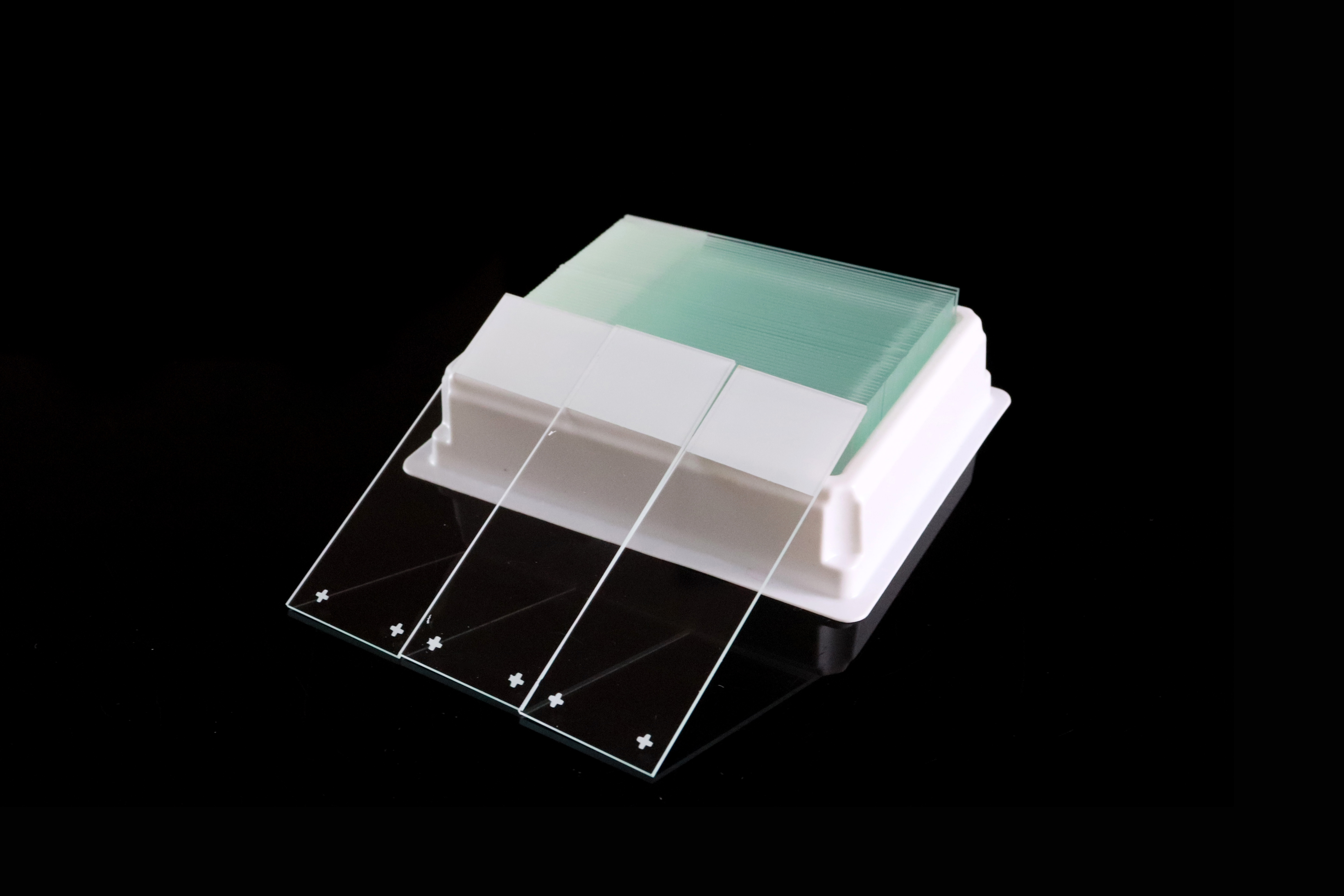
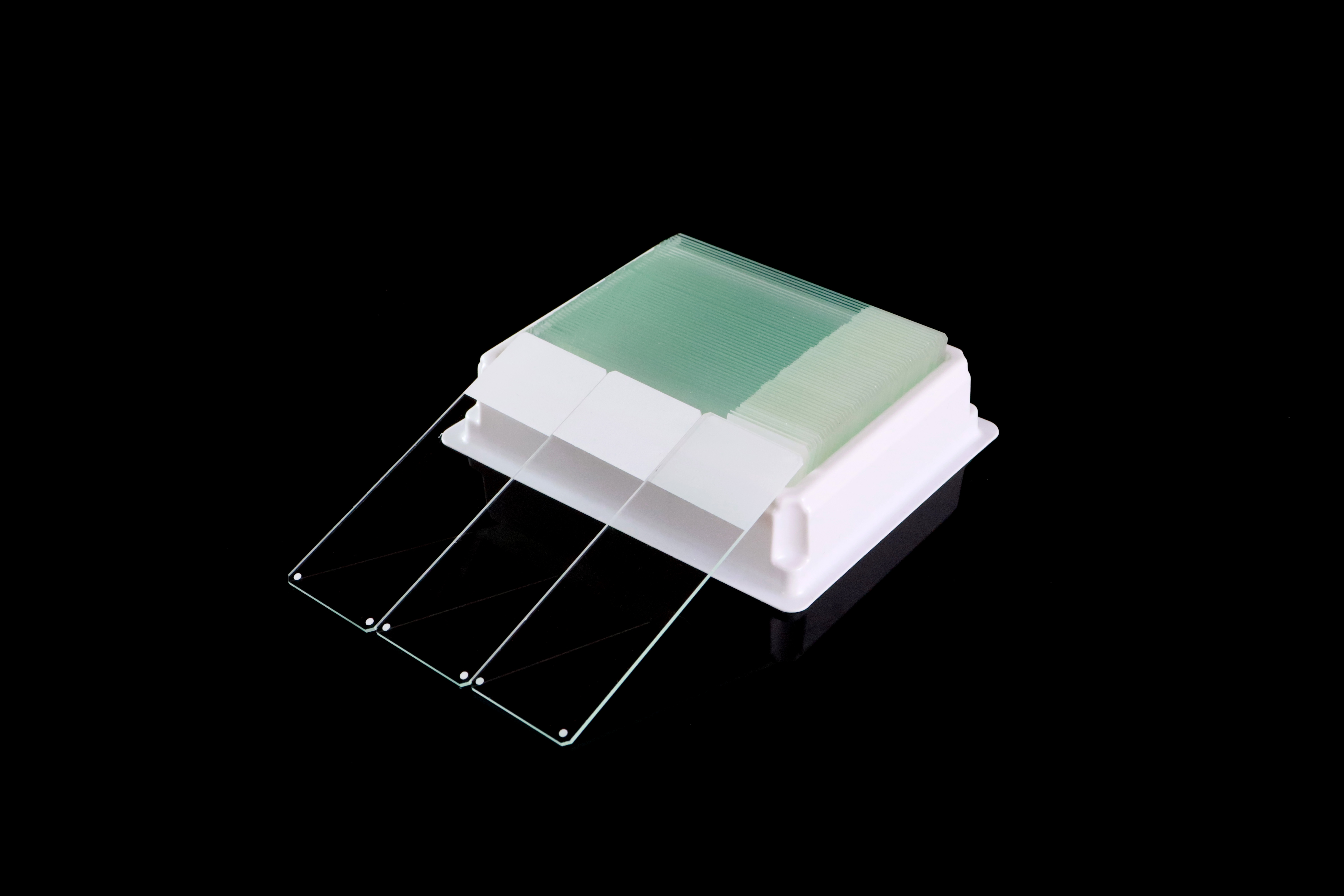
Microscope Slide vs. Coverslip vs. Prepared Slide
Item | Purpose | Purchasing distinction |
|---|---|---|
Blank microscope slide | Supports a specimen prepared by the user | Select the glass, surface, marking area and format |
Cover slip or cover glass | Covers the specimen in methods that require it | Select the size and thickness appropriate to the preparation and optics |
Prepared microscope slide | Already contains a mounted specimen | Select the specimen, preparation quality and intended teaching or reference use |
Not every preparation requires a coverslip. The specimen method and microscope objective determine the appropriate arrangement. Coverslip thickness can affect image quality with objectives designed for a specified cover glass, so it should be checked separately from slide thickness. croscope Slides for Your Application
1. Define the Specimen and Preparation Method
State whether the slide will carry a tissue section, cell preparation, blood smear, microbiological specimen or teaching sample. Include the staining or imaging method so the supplier can identify the relevant product range.
2. Decide Whether Enhanced Adhesion Is Needed
Consider the processing conditions and existing experience with specimen retention. Where a charged or adhesive surface is required, request an exact model and evaluate samples in the intended workflow.
3. Specify the Identification Area
Confirm whether the laboratory uses handwriting, direct slide printing or attached labels. Specify the marking area’s size, side and color, then check compatibility with the actual marker, printer or label system.
4. Check Dimensions and Instrument Requirements
Specify length, width, thickness, tolerances, edge finish and corner geometry. These features matter for racks, printers, stainers, coverslippers and scanners. Nominally similar slides should not be assumed interchangeable in every instrument.
5. Approve Samples Before a Bulk Order
Evaluate the proposed catalog number against the laboratory’s requirements. Record the manufacturer, model and lot number, together with the assessed preparation, staining and equipment performance. Use the approved specification for repeat orders.
What Should Laboratories and Bulk Buyers Check?
Laboratories, distributors and importers should compare the complete specification alongside price. Useful checks include:
Glass and surface condition: Clarity, cleanliness, scratches and other visible defects.
Dimensions and consistency: Length, width, thickness and agreed tolerances.
Edges and corners: The specified finish, chipping and suitability for handling and equipment.
Marking performance: Frosted-area consistency and readability after relevant processing.
Surface treatment: The correct coating, intended application and sample-evaluation results.
Packaging: Slides per box, protection against breakage, labeling and carton quantities.
Traceability: Product reference, batch identification and relevant product documents.
Supply terms: Order quantity, sample availability, lead time and the specification for repeat purchases.
Slide quality is one part of microscopy performance. Specimen preparation, staining, mounting, optical setup and equipment condition also influence results. When a problem occurs, assess the full workflow rather than attributing every image or staining defect to the glass.
Frequently Asked Questions
What Is the Main Purpose of a Microscope Slide?
A microscope slide supports and positions a specimen for preparation and microscopic examination. It can also provide a place for specimen identification and support later storage or review.
What Are Microscope Slides Used for in Biology?
Biology laboratories use slides to observe cells, tissues, plant material, microorganisms and other specimens. The appropriate slide depends on the specimen, preparation method and imaging technique.
What Are Microscope Slides Used for in Pathology?
Pathology laboratories use slides to support tissue sections and cellular preparations for staining and examination. The selected slide must meet the laboratory’s requirements for identification, specimen retention and processing.
What Are Positive Charged Microscope Slides Used For?
Positively charged slides are designed to promote specimen attachment in specified applications. They are commonly evaluated for tissue sections and IHC workflows where retention during processing is important. The exact model should be assessed with the laboratory method.
Are Frosted Microscope Slides Adhesive?
A frosted marking area does not establish that the specimen surface is adhesive. Some products combine a frosted end with a charged or adhesive surface, so check both features in the product specification.
Do All Microscope Slides Need a Cover slip?
No. Cover slip use depends on the specimen preparation and microscope optics. Some methods examine uncovered preparations; others require a cover slip of a specified size and thickness.
Can One Slide Type Be Used for Every Laboratory Application?
No single specification should be assumed suitable for every method. Surface treatment, marking area, glass dimensions and instrument requirements can differ. Choose and evaluate slides for the intended application.
Microscope Slides from Chuangxin Medlab
Yancheng Chuangxin Medlab Co., Ltd. manufactures microscope slides for laboratory users, distributors, importers and OEM buyers. Our range includes:
Plain microscope slides for routine preparation and general microscopy.
Frosted microscope slides with dedicated identification areas.
Color-frosted microscope slides for marking and laboratory organization.
Positively charged microscope slides for applications requiring enhanced attachment.
Adhesive microscope slides for specified specimen-retention requirements.
Custom specifications and private-label packaging are available on request. Explore our microscope slide range to compare the available options. Chuangxin Medlab
To request a quotation or evaluation samples, share your specimen and application, slide dimensions, edge and corner requirements, marking area, required surface treatment, packaging and order quantity. For automated workflows, include the instrument model so the proposed specification can be checked before ordering.