

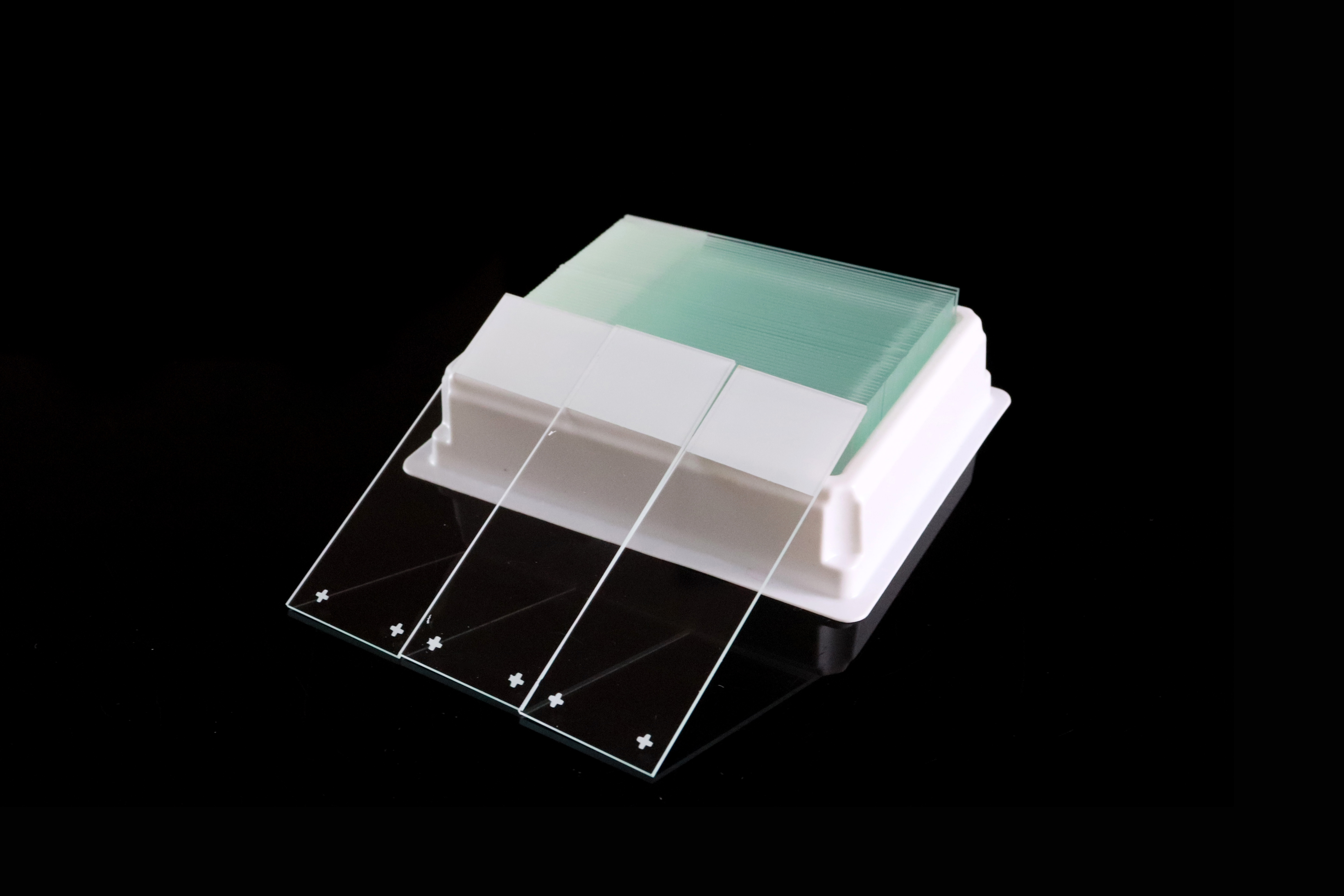
Microscope Slides for IHC: A Selection and Evaluation Guide
Selecting microscope slides for immunohistochemistry (IHC) involves more than finding a surface that holds tissue. The slide must support the intended preparation, retrieval and staining workflow while allowing the laboratory to obtain acceptable staining results.
For pathology laboratories, distributors and importers, a useful selection process combines a defined product specification with application testing. This guide explains what to specify, what to compare and how to record the results before bulk purchasing.
Quick Answer: Which Microscope Slides Are Suitable for IHC?
IHC commonly uses positively charged or other adhesion-treated microscope slides selected for the particular assay and laboratory workflow. Evaluate tissue retention together with staining intensity, expected localization, background, morphology and equipment compatibility.
Start with the assay, instrument and slide manufacturers' instructions. Then compare the proposed product with the laboratory's currently approved slide under representative conditions. A claim of strong adhesion alone is insufficient to establish IHC suitability.
Why Tissue Retention and Staining Quality Must Be Assessed Together
Depending on the method, IHC may involve heat-induced epitope retrieval (HIER), enzymatic pretreatment, repeated washes and automated reagent application. These conditions make the interaction between the section and the slide important.
Useful tissue retention means preserving the section for assessment. It should not be judged only by whether some tissue remains at the end.
A slide can retain tissue yet produce unacceptable staining, while staining differences can also arise from fixation, reagents, retrieval or other preparation variables. Evaluate retention and staining as separate outcomes, and investigate differences systematically.
Charged, Adhesive or Plain Slides: Where to Start
Positively Charged Slides
Positively charged microscope slides have a surface treatment intended to improve the attachment of tissue sections and cells. They are commonly considered for IHC workflows that require enhanced adhesion.
However, products described as positively charged can differ in surface properties and performance. Select the exact product according to its intended use and laboratory evaluation.
Other Adhesion-Treated Slides
“Adhesive” is a broad product description. Some adhesive slides also have a positively charged surface, so these terms can overlap.
Ask for the product's intended applications, preparation instructions and relevant performance information. Hydrophilic surface properties may support wetting by aqueous reagents, but a wettability claim does not establish complete IHC performance.
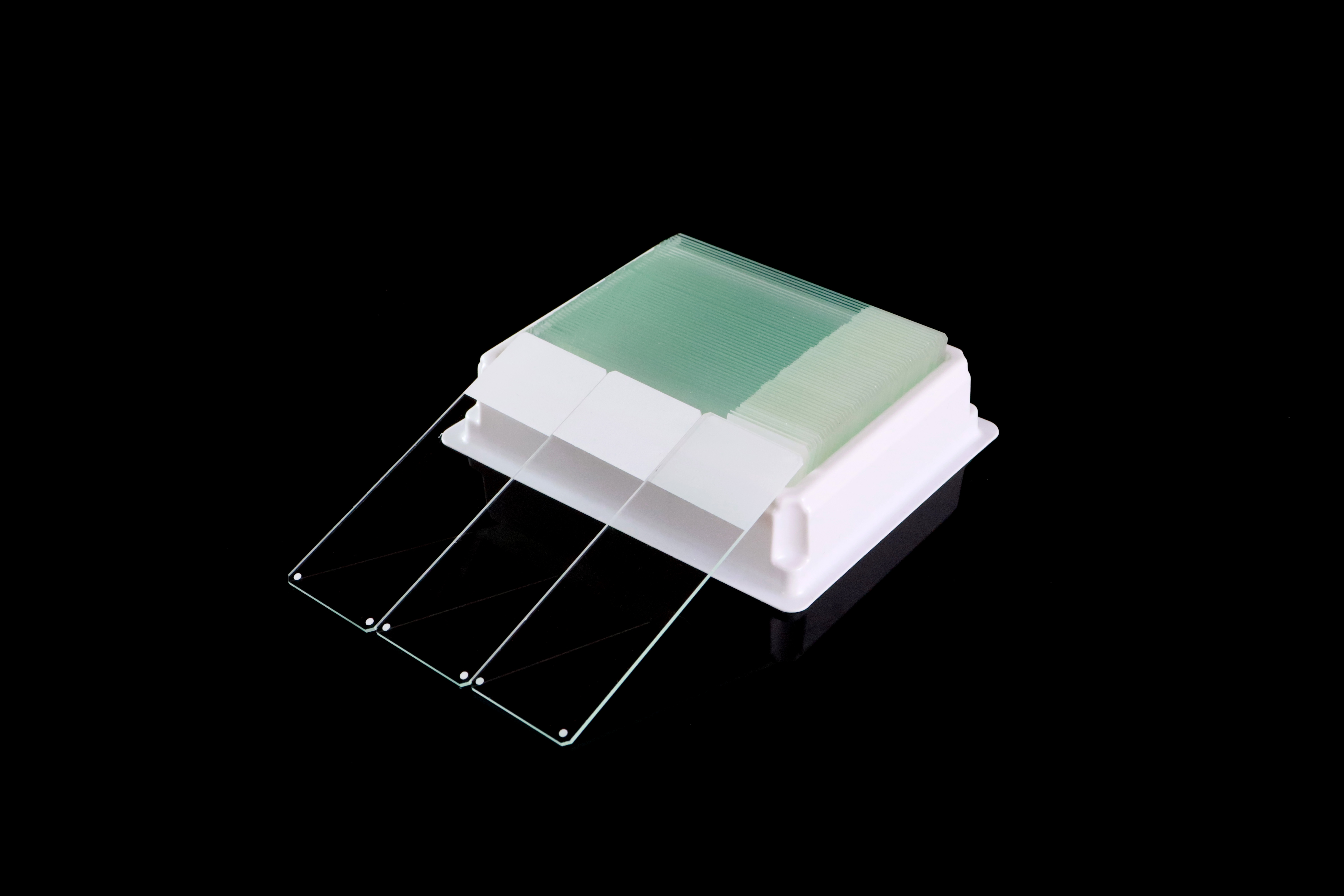
Plain Slides
Plain, untreated slides do not provide the same specified adhesion treatment. Their use should be supported by the intended assay and the laboratory's established process.
A slide that works for routine H&E staining should not automatically be treated as qualified for an IHC protocol with different retrieval and washing conditions.

IHC Microscope Slide Selection Checklist
Selection factor | What to specify or evaluate |
|---|---|
Specimen preparation | Formalin-fixed, paraffin-embedded (FFPE), frozen or another intended preparation |
Tissue requirements | Routine tissues and representative samples with a history of lifting or loss |
Surface treatment | Exact product and coating description, intended use and preparation instructions |
Retrieval conditions | Relevant method, buffer, temperature and duration from the assay protocol |
Staining performance | Expected signal, localization, background and tissue morphology |
Reagent coverage | Suitability of surface wetting for the intended application system |
Physical dimensions | Length, width, thickness, tolerances, edges and corners |
Identification | Printable area or labels, barcode readability and resistance to processing |
Storage and supply | Labeled storage conditions, expiry, packaging, lot identification and repeat-order consistency |
Use the checklist to define the candidate product before requesting samples. It also helps distinguish a general sales sample from the configuration intended for the order.
Evaluate Compatibility with the Complete Workflow
Check the requirements of the exact automated stainer and any associated printer, rack, retrieval system or scanner. A brand-family description is less useful than a full equipment model and the applicable slide requirements.
For manual staining, assess slide handling and reagent coverage within the laboratory's established method. A surface description such as “hydrophilic” should be evaluated in that process rather than used as a universal ranking of slide quality.
Confirm that printing and tissue placement can coexist within the permitted marking and staining areas. Check identification readability after processing as well as immediately after printing.
Where an assay specifies particular slide products, follow the assay instructions when planning the evaluation.
How to Evaluate IHC Slide Samples
1. Define the Comparison Before Testing
Identify the currently approved reference slide and the candidate product. Record model, lot, surface treatment, expiry and storage history.
Select representative tissues and assays, including relevant difficult specimens and controls capable of revealing changes in performance. For example, a laboratory using tissue microarrays may include core retention in its evaluation.
Set acceptance criteria before the trial. The laboratory's responsible staff should define what constitutes acceptable retention, staining and reproducibility.
2. Use Comparable Sections and Document Preparation
Where practical, compare sections from the same tissue blocks, recognizing that serial sections can differ.
Keep preparation and staining conditions comparable. Record section thickness, mounting, drying, time before staining and the complete assay conditions. If a product requires a different preparation under its instructions, document that difference explicitly.
Follow the relevant instructions for drying and baking. Increasing heat or extending baking time is not a universal solution to tissue loss.
3. Compare Through the Intended IHC Procedure
Assess the candidate in the actual retrieval and staining workflow being considered. Keep the reference comparison identifiable throughout the run.
Include the positive, negative and reagent controls required by the assay and laboratory procedures. Have qualified staff review expected staining intensity and localization, including controls that can reveal reduced sensitivity.
A result from one successful stain should not be extended automatically to other assays, tissue preparations or platforms.
4. Record Retention, Staining and Background Separately
Inspect the whole relevant section for loss, lifting, folds and damage. Also assess expected staining and morphology.
Look for unexpected staining in the tissue and on exposed glass. Some slide surfaces or added adhesives can contribute to background, but background alone does not identify its cause. Compare the controls and reference slide before attributing a problem to the coating.
For fluorescence-based methods, include an assessment of slide-related background in the relevant detection channels.
5. Confirm Repeatability and Approve the Defined Use
Choose repeat runs, sample numbers and lot coverage according to the intended use and the laboratory's evaluation procedures.
Retain images, observations, preparation details and the purchasing specification. Record the conditions under which the slide performed acceptably.
Before routine introduction, complete the laboratory's applicable verification and change-control process. For subsequent orders, agree on incoming checks and notification of product changes.
A Practical IHC Slide Evaluation Record
Outcome | Suggested record |
|---|---|
Tissue retention | Location and extent of loss or lifting; number of sections affected |
Staining performance | Expected signal intensity and localization compared with the reference |
Background | Unexpected deposits or staining in tissue and on exposed glass |
Morphology | Section integrity and features relevant to interpretation |
Workflow performance | Handling, instrument fit, reagent coverage and identification readability |
Reproducibility | Results by run and lot, including recorded preparation differences |
Document the number examined alongside any failures. Avoid presenting a small convenience sample as a reliable failure rate for the entire shipment.
Storage, Handling and Repeat Supply
Follow the slide manufacturer's storage, handling and expiry instructions. Keep unused slides protected as directed and avoid touching the specimen surface.
Distinguish the storage life of unused slides from the acceptable storage of cut tissue sections. Section age and storage requirements also depend on the tissue and assay; use the relevant instructions.
For bulk purchasing, retain lot identification and agree on how changes to the coating, glass, packaging or specification will be communicated. Our microscope slide quality inspection guide covers general incoming checks.
What Should Buyers Send with an IHC Slide Inquiry?
Provide the supplier with the intended tissue preparations, assay types, retrieval conditions and equipment models. Add the required dimensions, marking area, surface treatment, packaging and order volume.
Request samples matching the proposed production specification, relevant product instructions, lot information and available application data. Define sample acceptance with the laboratory that will use the slides.
For distributors, keep customer approvals linked to the particular slide and workflow tested.
Frequently Asked Questions
Are positively charged slides and adhesive slides different categories?
The descriptions can overlap. A positively charged slide may also be described as adhesive. Compare the exact surface treatment, intended use and evaluation results.
Does stronger tissue adhesion always mean better IHC performance?
No. The slide must also support acceptable staining, background, morphology and workflow performance. Evaluate these outcomes separately.
Can a slide approved for H&E be used for IHC?
It needs assessment for the intended IHC method. Retrieval, reagents and processing conditions may differ from the H&E workflow.
Are all IHC slides compatible with every automated stainer?
No. Confirm the exact instrument and assay requirements, physical specifications and application performance. Qualification on one combination does not establish suitability for all systems.
How many samples should buyers test?
There is no single sample count suitable for every purchase. Base the plan on intended assays, tissue variety, production lots, workload and the laboratory's evaluation requirements.
Discuss IHC Slide Requirements with Chuangxin Medlab
Chuangxin Medlab manufactures microscope slides for laboratories, pathology suppliers, distributors, importers and OEM customers. Our range includes positively charged microscope slides and adhesive microscope slides.
Share your tissue preparation, IHC workflow, equipment, slide specification and packaging needs so the proposed product can be defined for sample evaluation.
Contact Chuangxin Medlab to discuss product specifications, sample availability, OEM options and a bulk quotation.