






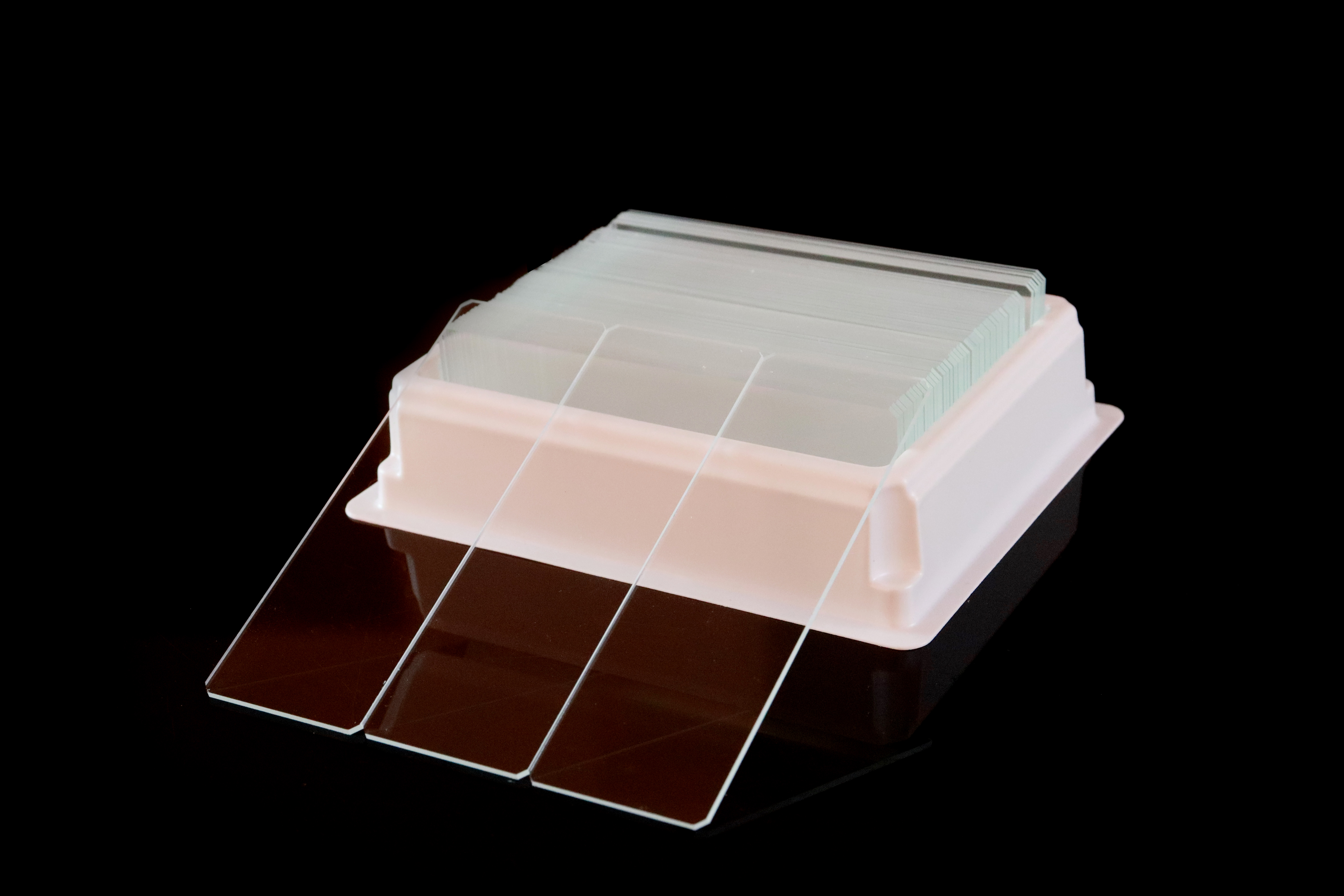
What Are the Main Types of Microscope Slides?
The main types of microscope slides are plain, single frosted, double or twin frosted, color frosted, positively charged, adhesive, concave, and specialized slides. These names describe different features: labeling areas, specimen adhesion, slide geometry, or a particular application. A single slide can combine several features, such as a colored marking area, a charged specimen surface, and ground edges.
For routine observation, laboratories commonly consider plain slides. A frosted or colored marking area helps identify specimens. Charged or adhesive surfaces address specimen retention, while concave slides provide space for selected liquid or thicker preparations. The appropriate choice depends on the laboratory method and the exact product specification.
This guide explains how the categories differ, which specifications matter, and how to prepare a clear purchase request for laboratory or OEM supply.
Microscope Slide Types: Quick Comparison
Slide type | Defining feature | Common reason to select it | Important purchase check |
|---|---|---|---|
Clear glass without a dedicated frosted marking area | Routine observation, teaching, and general specimen preparation | Dimensions, cleanliness, edge finish, and identification method | |
A frosted marking area at one end in the specified face configuration | Handwritten specimen identification | Marking area size, face, and resistance of the chosen writing method to processing | |
Two marking areas or a marking area on both faces, depending on the reference | A particular labeling or slide orientation requirement | Exact position and number of frosted areas; request a drawing | |
A colored marking field | Visual classification and specimen identification | Color, usable print area, and compatibility with the actual printer and ribbon | |
A surface designed to carry a positive charge | Tissue retention in appropriate histology methods | Intended use, surface treatment, storage conditions, and method validation | |
A surface treatment designed to improve specimen attachment | Preparations that need additional adhesion | Coating identity and performance in the intended procedure | |
One or more depressions in the glass | Selected wet preparations or specimens needing extra space | Cavity diameter, depth, and suitability for the observation method | |
Specialized slide or chamber | A grid, reaction well layout, defined chamber depth, or other application-specific feature | Cell counting or a defined assay | Measurement geometry, material, coverslip requirements, and manufacturer instructions |
Charged slides can also be adhesive slides. A frosted or colored marking field can appear on either category. The table compares features; it does not imply that every product belongs to only one group.

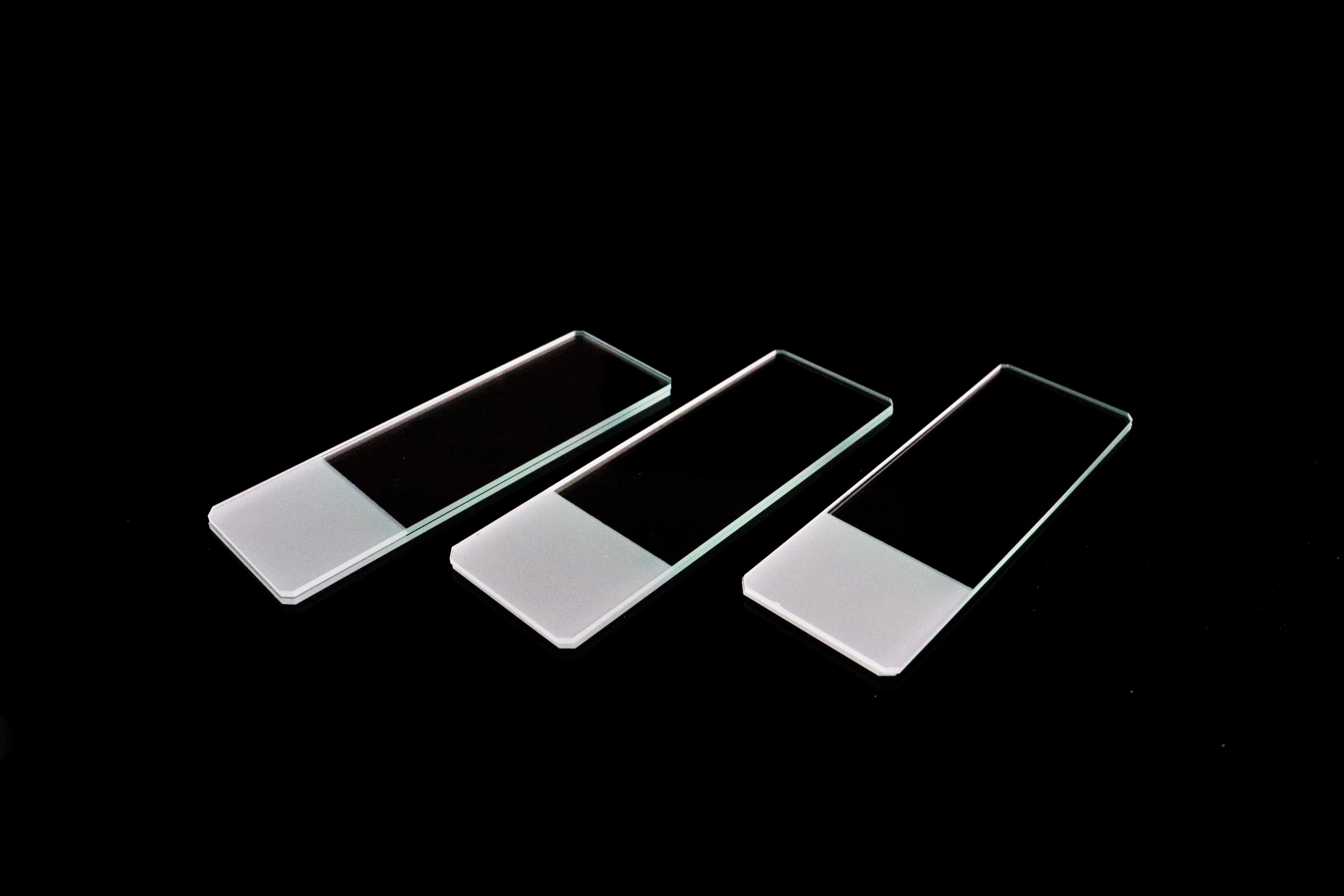

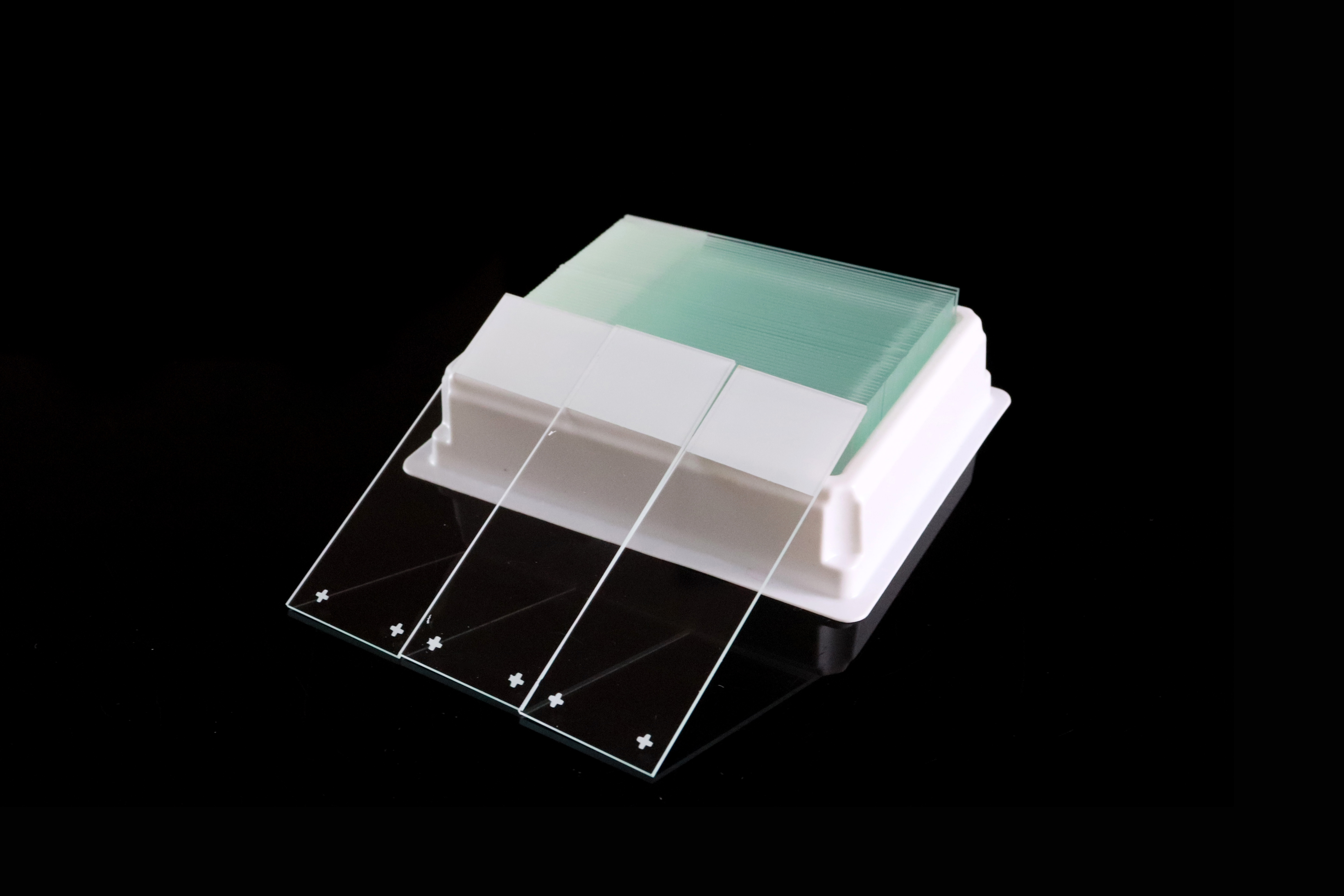
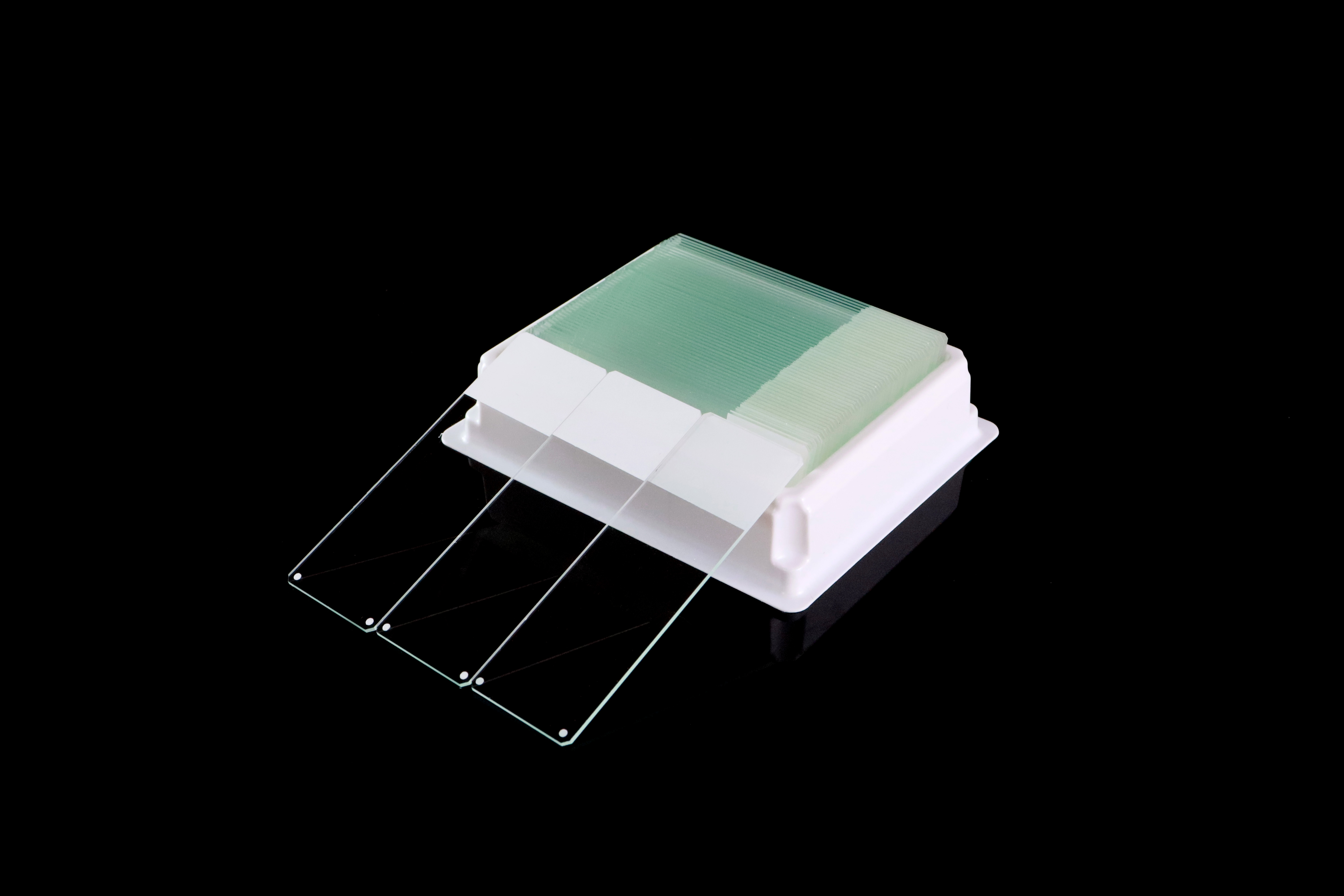
Understand Four Separate Slide Features Before Ordering
Selecting a microscope slide becomes easier when you describe four features separately:
Identification: plain, frosted, or color frosted; marking area position and dimensions.
Specimen surface: untreated glass, positively charged treatment, or another adhesive coating.
Geometry: flat glass, a cavity, reaction wells, or a defined counting chamber.
Mechanical specification: length, width, thickness, edge finish, and corner configuration.
For example, “color frosted slide” identifies the marking field but does not tell a buyer whether the specimen surface is charged. Likewise, “ground edge” describes the edge finish and does not establish tissue adhesion or printer compatibility.
Write all relevant features into the purchase specification. This reduces uncertainty when comparing similar catalog names from different suppliers.
Plain and Frosted Microscope Slides: What Is the Difference?
Plain Microscope Slides
Plain microscope slides provide a clear glass surface without a dedicated frosted labeling field. They are useful for general observation and specimen preparation when the method does not call for a particular adhesive surface.
Plain slides still need a reliable identification method. Depending on the workflow, a laboratory may use a suitable label or another approved marking method. The absence of frosting does not mean that a specimen cannot be identified permanently.
For procurement, check the glass specification, dimensions, surface condition, and edge finish. Do not assume that all plain slides have identical thicknesses or handling characteristics.
Single Frosted Microscope Slides
Single frosted slides have a marking field at one end. Its purpose is to provide space for specimen information while leaving the observation area available for the preparation.
The order should state which face is frosted and how large the marking field must be. A handwritten label also needs to remain legible through the intended handling, staining, and storage conditions.
Chuangxin Medlab's 7105 single frosted microscope slides are currently listed with a single frosted end on one face and ground edges. The 7105-1 reference is listed with cut edges. These are product-specific configurations, rather than a universal definition of every frosted slide.
Double and Twin Frosted Microscope Slides
“Double frosted” and “twin frosted” require a closer specification check. Catalog terminology may refer to marking areas on both faces or to marking fields at two ends.
For example, Marienfeld's frosted slide reference describes a marking area on both faces. Chuangxin Medlab's current 7107 twin frosted product page describes two frosted ends on one face.
Ask for a product drawing showing the marking areas, faces, and remaining specimen area. More frosting does not, by itself, improve specimen adhesion.

Color Frosted Microscope Slides
Color frosted slides use a colored marking field to support specimen identification and visual organization. Laboratories may assign colors to departments, preparation categories, or internal workflows.
The color system should follow the laboratory's own identification procedure. There is no universal meaning for each color.
If the field will be printed, test it with the specified printer model, ribbon or ink, and processing conditions. A color frosted surface alone does not establish compatibility with every slide printer.
Positive Charged vs. Adhesive Microscope Slides
What Is a Positive Charged Microscope Slide?
A positively charged microscope slide has a treated surface intended to help retain specimens such as tissue sections. Manufacturer instructions determine which preparations and processing conditions the selected reference supports.
These slides are commonly considered for histology workflows in which specimen attachment matters. For example, Marienfeld lists its HistoBond+ slides for immunohistochemistry and in situ hybridization and identifies positive charge as the mechanism supporting adhesion.
The marking field and the charged specimen surface serve separate purposes. A frosted end helps identify the slide; the adhesion treatment concerns the working surface.
What Is an Adhesive Microscope Slide?
An adhesive slide is designed to improve specimen attachment through a specified surface treatment. The term describes the intended function, while “positively charged” describes a surface characteristic. Therefore, the two categories can overlap.
Manufacturers illustrate this overlap directly: HistoBond+ adhesive slides use a positively charged surface, and Leica Apex adhesive slides have a proprietary coating that provides a positive charge.
Ask the supplier to identify the coating or treatment and its intended use. “Adhesive,” “charged,” and “hydrophilic” do not provide an interchangeable description of the coating chemistry. Hydrophilicity describes how a surface interacts with water; it is not, by itself, the name of an adhesive formulation.
How Should Buyers Compare Adhesion Products?
Compare the exact slide reference in the complete intended workflow. Relevant questions include whether the preparation stays attached, whether staining remains acceptable, and whether the slide works with the laboratory's labeling and handling equipment.
For methods involving antigen retrieval or other demanding processing steps, follow the reagent, equipment, and slide manufacturers' instructions and the laboratory's validated method. There is no single coating that can be declared the best for every specimen and procedure.
Concave Microscope Slides for Wet Preparations
Concave slides, also called cavity or depression slides, contain a recessed area that creates additional space for a preparation. They can be considered for selected liquid specimens or observations where a flat slide provides insufficient space.
The cavity is a physical feature, rather than an adhesion treatment. A slide may have one or more cavities, and the number, diameter, and depth vary by product. Marienfeld's cavity slide catalog, for example, lists several configurations.
Specify the cavity geometry and confirm that it suits the specimen, microscope, and intended preparation method. A deeper recess is not automatically more suitable: the optical arrangement and observation procedure also matter.
An ordinary cavity slide does not provide a defined counting volume simply because it holds a liquid. Quantitative counting requires the appropriate measurement geometry and instructions.
Specialized Microscope Slides and Counting Chambers
Counting Chambers and Hemocytometers
A counting chamber combines a defined counting grid with a specified chamber depth. Together, these establish the observation volume used in the counting method. The correct chamber cover glass is also part of the measurement setup.
For example, Marienfeld lists the standard Neubauer-improved grid with a 0.1 mm chamber depth. Other chamber designs use different grids and depths, so that figure should not be applied to every counting chamber.
When sourcing a hemocytometer, specify the grid type, depth, cover glass requirements, and intended method. Do not substitute a regular slide or an uncalibrated cavity slide on the basis of similar appearance.
Reaction Well and Other Application-Specific Slides
Some specialized slides use defined reaction areas to organize multiple preparations on one slide. These areas can be formed by a printed mask rather than by depressions cut into the glass.
Check the number, diameter, and spacing of the wells, along with the surface treatment and resistance of the mask to the intended reagents. Marienfeld's reaction well slides are one example of this category.
Other assays may call for a particular substrate, grid, coating, or optical property. Purchase those products against the method's specification instead of treating every specialized slide as a substitute for general glass slides.
How to Choose a Microscope Slide for Your Application
Start with the preparation method, then add identification and equipment requirements. The following table is a starting point for evaluating products; the final selection depends on the validated laboratory procedure.
Laboratory requirement | Slide feature to evaluate | What to confirm before purchase |
|---|---|---|
General observation or teaching | Plain glass slide | Size, surface cleanliness, edges, and specimen identification |
Handwritten specimen identification | A specified frosted marking field | Field position and dimensions; legibility after processing |
Color-based specimen organization | Color frosted field | Internal color convention and identification procedure |
Tissue retention during demanding processing | A suitable charged or adhesive surface | Performance in the complete staining or assay method |
Wet preparation needing additional space | An appropriate cavity | Cavity geometry and observation requirements |
Quantitative cell or particle counting | A defined counting chamber | Grid, chamber depth, and compatible cover glass |
Automated printing or handling | An equipment-compatible slide reference | Manufacturer specifications and a trial using the actual system |
If a routine slide is already performing satisfactorily, identify the specific requirement before changing it. A different color, coating, thickness, or edge configuration can affect more than one part of the workflow.
Microscope Slide Sizes, Thicknesses, Materials, and Edges
Common Nominal Dimensions
Common catalog dimensions include the following sizes, expressed here as width × length:
Nominal size | Meaning for purchasing |
|---|---|
25 × 75 mm | A common metric slide format |
25.4 × 76.2 mm | Exactly 1 × 3 inches |
26 × 76 mm | Another frequently listed metric format |
25 × 75 mm is not exactly the same size as 1 × 3 inches. Do not use the descriptions interchangeably in a purchase order. Confirm the exact nominal dimensions and permitted tolerances, particularly for automated equipment.
Thickness and Glass Specification
Thickness depends on the product reference. Chuangxin Medlab currently lists 1.0–1.2 mm for several of its standard plain and frosted models. This range should not be applied automatically to specialty slides, cavity slides, or products from other manufacturers.
Ask for the required thickness and tolerance as separate specifications. Also confirm the glass composition or grade, the surface condition, and any optical requirements of the application.
Soda-lime glass is listed for multiple standard slide products. Descriptions such as “super white” are commercial grade names and should be supported by the supplier's specification. Do not infer that a slide uses borosilicate glass merely because the same supplier offers borosilicate cover glass.
Cut Edges, Ground Edges, and Corner Configuration
Cut edges and ground edges describe different finishes. Ground edges undergo additional finishing, while cut edges require handling appropriate to their condition. Inspect samples for chips, irregularities, and consistency.
Corner configuration is another specification. A 45° corner treatment and a 90° corner should be identified separately from the edge finish.
For automated systems, evaluate the complete slide: dimensions, thickness, flatness, corners, marking field, and surface treatment. “Ground edge” alone does not establish compatibility with a printer, stainer, coverslipper, or scanner.
Microscope Slides vs. Cover Glass: What Is the Difference?
A microscope slide supports the specimen. Cover glass, also called a coverslip, is a thinner piece placed over selected preparations according to the observation method.
The two products have different dimensions and requirements. Cover glass selection can depend on the microscope objective, mounting method, and specimen preparation. A counting chamber may require a particular chamber cover glass rather than an ordinary coverslip.
Order slides and coverslips as separate specifications. Chuangxin Medlab's microscope cover glass catalog lists different shapes and product categories for buyers to evaluate.
Chuangxin Medlab Microscope Slide Options for Laboratory and OEM Buyers
Yancheng Chuangxin Medlab Co., Ltd. is a China-based manufacturer supplying microscope slides under the S.D.T.C. brand. Its published catalog includes standard glass slides, marking-field options, positively charged slides, adhesive slides, and related microscopy products.
Published model or category | Catalog description | Main specification to discuss |
|---|---|---|
Plain slides; 7101 ground edge, 7102 cut edge | Glass, dimensions, thickness, and edges | |
Single frosted slides; ground-edge and cut-edge references | Marking field layout and writing requirements | |
Twin or double frosted references; ground-edge and cut-edge versions | A drawing of the marking fields, faces, and usable specimen area | |
Colored marking field | Color, field dimensions, and printer requirements | |
Positive-charge surface treatment | Intended method and reference-specific documentation | |
Adhesion-oriented slide category | Surface treatment, storage conditions, and sample evaluation | |
Cavity slide category | Cavity number, diameter, and depth | |
Counting chamber category | Grid, chamber depth, and cover glass specification |
Chuangxin Medlab also lists OEM and private-label services. Confirm the actual customization scope, order quantity, packaging, documentation, and delivery terms in the quotation for the selected product.
What to Include in a Microscope Slide Purchase Request
A clear request helps the manufacturer quote the intended product and provide suitable samples. Include:
Application: specimen type, preparation method, and relevant processing conditions.
Dimensions: nominal width, length, thickness, and required tolerances.
Glass and mechanical finish: material specification, cut or ground edges, and corner configuration.
Identification field: color, size, position, face, and handwriting or printer requirements.
Specimen surface: untreated, positively charged, or a specified adhesive treatment.
Special geometry: cavity dimensions, reaction well layout, or counting chamber requirements.
Packaging: slides per box, boxes per carton, labeling, and any private-label artwork.
Commercial requirements: sample quantity, planned order volume, destination, and required delivery date.
For a replacement product, send the current slide's specification or a clear drawing as well as its catalog name. A model number alone may not capture the configuration supplied by another manufacturer.
To discuss the published range, contact Chuangxin Medlab for a quotation and sample availability. Share your application and equipment requirements so the proposed slide can be evaluated before a bulk order.
Frequently Asked Questions About Microscope Slide Types
What Are the Most Common Types of Microscope Slides?
Common categories include plain, frosted, color frosted, positively charged, adhesive, and concave slides. Specialized products include reaction well slides and counting chambers. These categories can overlap because labeling, adhesion, and geometry describe different features.
Are Frosted Microscope Slides the Same as Charged Slides?
No. Frosting identifies a marking area used for specimen information. A charged treatment concerns the specimen surface and its adhesion properties. A particular slide can have both features, but a frosted marking area alone does not establish that the working surface is charged.
Are Positive Charged Slides Also Adhesive Slides?
They can be. Positive charge is one way manufacturers provide an adhesion-oriented surface. The terms are not mutually exclusive. Check the exact treatment and intended use instead of assuming that “charged” and “adhesive” always identify different products.
Does Double Frosted Mean Both Ends of the Slide Are Frosted?
Not necessarily. Published references use different marking layouts, including fields on both faces and fields at two ends. Request a drawing showing the location and dimensions of each field before ordering.
Is a 25 × 75 mm Slide the Same as a 1 × 3 Inch Slide?
No. One inch equals 25.4 mm, so a 1 × 3 inch slide measures 25.4 × 76.2 mm. State the intended dimensions explicitly when ordering and check the tolerance requirements of any automated equipment.
Which Microscope Slides Should Be Considered for IHC?
Evaluate charged or adhesive products that the manufacturer lists for the intended IHC method. Confirm specimen retention and staining performance in the complete laboratory workflow, including any antigen retrieval steps. The most suitable reference depends on the method and specimen.
Can Any Color Frosted Slide Be Used in a Slide Printer?
No universal compatibility should be assumed. The marking surface, field dimensions, slide geometry, and printing system must work together. Check the equipment manufacturer's requirements and test the selected slide with the actual printer, consumables, and processing conditions.
Can a Concave Slide Replace a Hemocytometer?
An ordinary concave slide cannot replace a counting chamber merely because both hold liquid. A hemocytometer uses a specified grid and chamber depth to establish a counting volume. Select the chamber and cover glass required by the counting method.

Related Articles
-
 Compare 10 microscope slide manufacturers worldwide in 2026, including Chuangxin Medlab. Explore plain, frosted, charged, and adhesive slides. Review model specifications, printer compatibility, pack quantities, OEM options, and sample checks to choose a manufacturer for your laboratory or business.
-
 Compare 7 microscope slide manufacturers and suppliers for Malaysia in 2026, including Chuangxin Medlab. Review local buying channels, plain and adhesive slides, dimensions, box quantities, OEM options and sample checks. Learn how to compare quotations and plan delivery to your Malaysian laboratory.
-
 Compare 5 microscope slide manufacturers and suppliers for Turkey in 2026, including Chuangxin Medlab. Review local purchasing channels, plain and adhesive slides, product specifications, box quantities and OEM options. Use the buying checklist to request samples and compare quotes for your own lab.
- Compare six microscope slide manufacturers and suppliers for Colombia, including local purchasing channels and Chuangxin Medlab. Explore product references, slide sizes, packaging, surface options and sample checks to prepare clear quotations for routine microscopy, pathology and OEM supply in 2026.
