






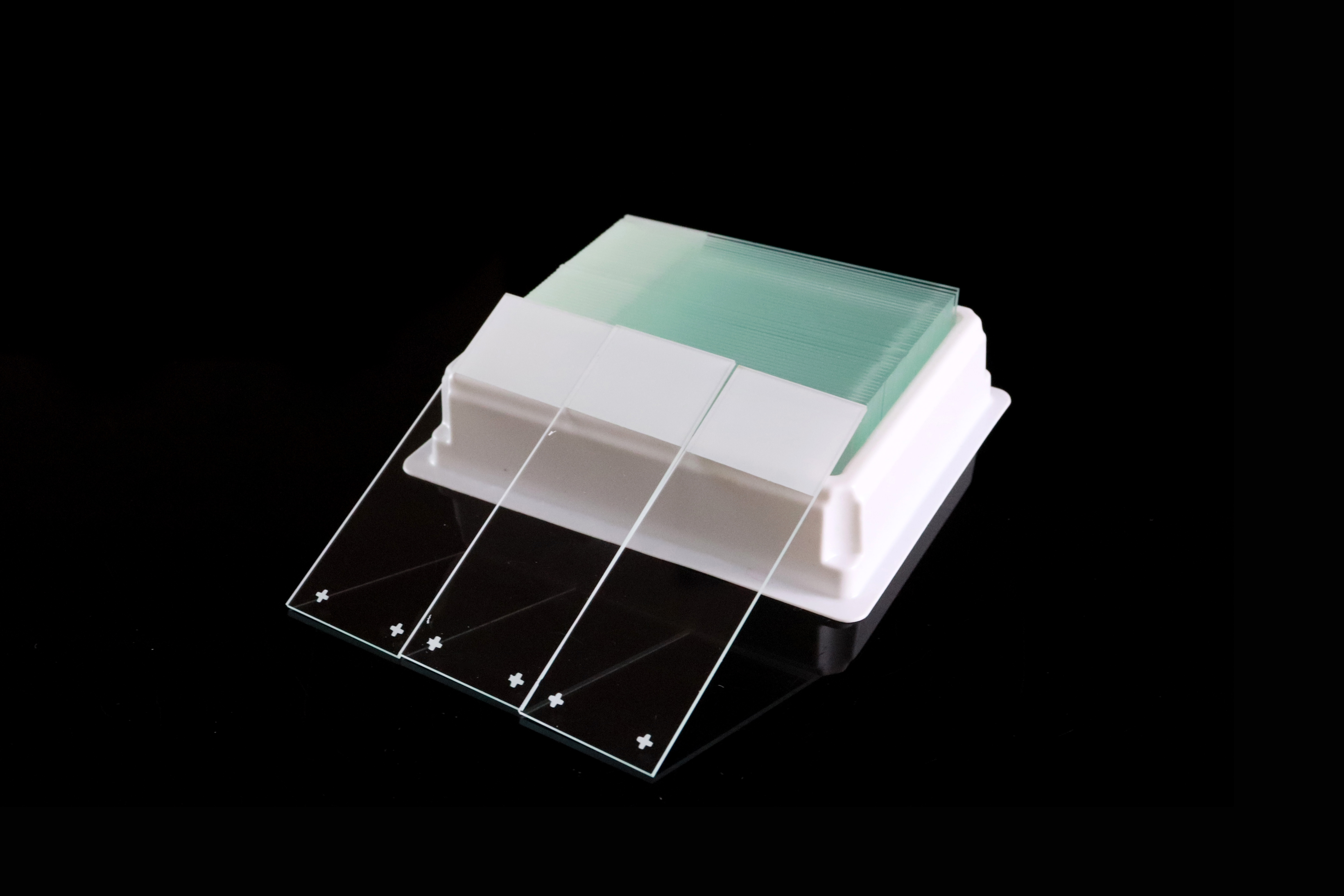
Positively Charged vs Adhesive Microscope Slides: How to Choose
When selecting microscope slides for histology or pathology, buyers often encounter two descriptions: positively charged and adhesive. The names can suggest separate product categories, yet a slide may carry both descriptions.
For laboratories, distributors and importers, the useful comparison is between specific products: their surface treatment, documented applications and performance in the intended workflow. This guide explains how the terms relate and what to evaluate before approving samples or placing a bulk order.
Quick Answer: What Is the Difference?
“Positively charged” describes a surface property used to promote specimen attachment. “Adhesive” is a broader description for slides designed to improve specimen adhesion. Positively charged slides are therefore one type of adhesion-enhancing slide, and an adhesive slide may also be positively charged.
Manufacturers may use these labels for different models within their own ranges. The names alone do not establish which product will retain tissue better, produce less background or work with a particular staining instrument.
Choose using the exact product specification and relevant sample evaluation.
What Are Positively Charged Microscope Slides?
Positively charged microscope slides have a treated surface designed to support the attachment of tissue sections or cells through electrostatic interactions.
The intended benefit is improved specimen retention during suitable laboratory processing. The result still depends on the particular slide, specimen preparation and procedure.
A product described as positively charged should be assessed against its manufacturer's information, including intended applications, handling instructions, storage requirements and any available performance data.
What Are Adhesive Microscope Slides?
Adhesive microscope slides are designed with a surface treatment that promotes specimen attachment.
The term describes the intended function and can cover different treatment systems. Some adhesive slides are positively charged; others may be described by a coating technology, proprietary treatment or application-specific product name.
When a supplier lists “positively charged” and “adhesive” as separate categories, ask what distinguishes the actual models. There may be differences in surface properties, intended use or available performance evidence, but those differences should be established from the product information.
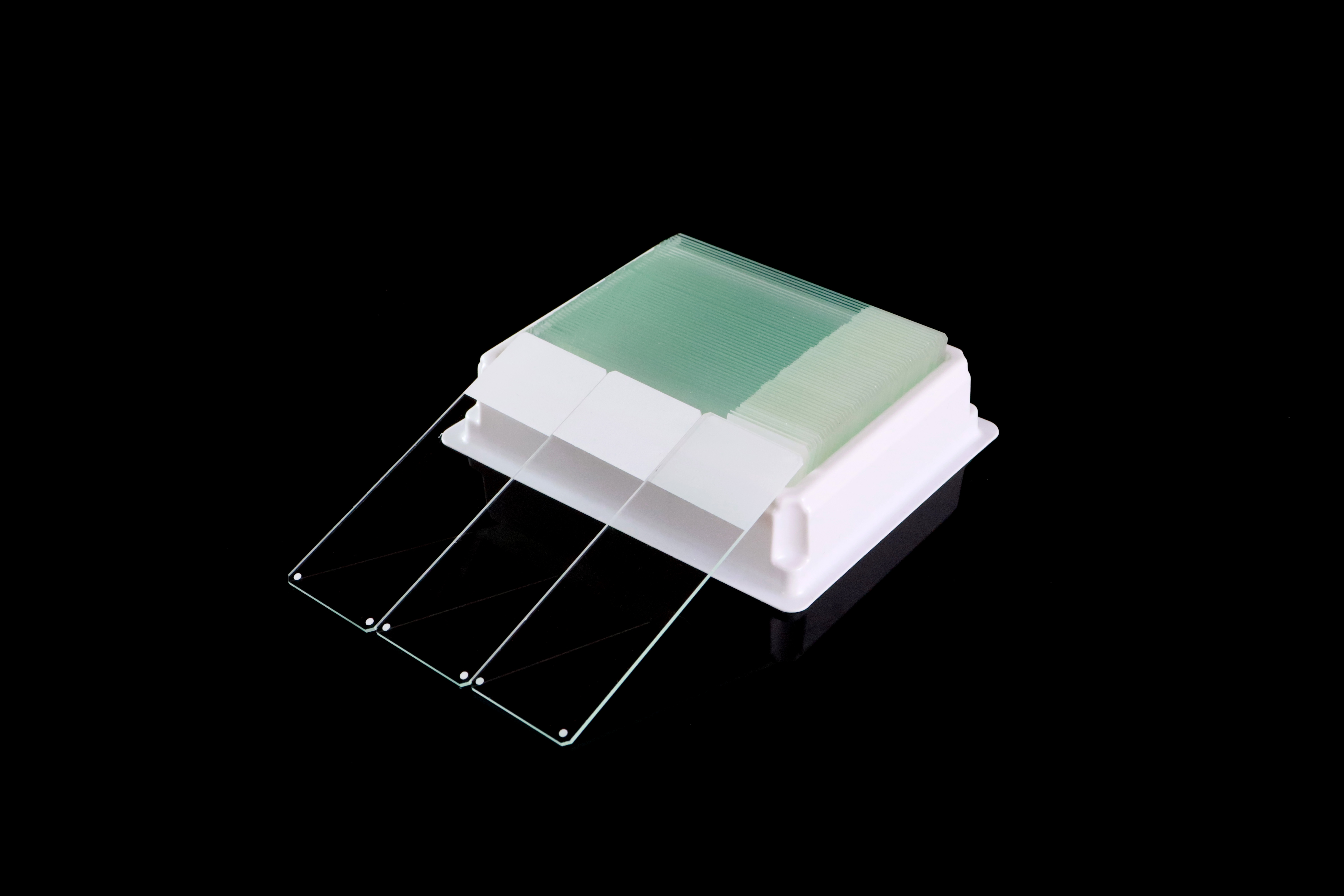
Positively Charged vs Adhesive Slides at a Glance
Aspect | Positively Charged Slides | Slides Labeled Adhesive |
|---|---|---|
What the name emphasizes | A positive surface charge | Improved specimen attachment |
Relationship between the terms | An adhesion-enhancing slide may be described this way | May include positively charged products |
Surface treatment details | Require information for the specific model | Require information for the specific model |
Application suitability | Depends on the product and laboratory workflow | Depends on the product and laboratory workflow |
Basis for choosing | Relevant performance evidence and sample evaluation | Relevant performance evidence and sample evaluation |
A category name is a starting point for product comparison. It is not a ranking of adhesion strength.
Charge, Hydrophilicity and Tissue Retention
These properties are related to surface performance but describe different things:
Surface charge concerns the electrical properties of the treated surface.
Hydrophilicity concerns how readily an aqueous liquid wets the surface.
Tissue retention concerns whether the specimen remains attached through the intended processing steps.
An adhesive slide can be both positively charged and hydrophilic. However, good wetting does not, by itself, demonstrate tissue retention throughout a staining procedure.
Where these properties matter, ask what the manufacturer has evaluated and compare the finished slides under the intended laboratory conditions.
How to Choose for Your Laboratory Workflow
Start with the application, specimen and equipment. Record the exact slide model currently used and the reason for considering an alternative.
Workflow | What to Evaluate |
|---|---|
Routine histology and H&E staining | Specimen retention, staining appearance and consistency in the established procedure |
IHC, including workflows with antigen retrieval | Retention through the complete procedure, background and relevant staining performance |
Automated staining | The specific instrument, protocol, slide format and usable specimen area |
Frozen sections or specimens prone to detachment | Performance with representative specimens under the laboratory's established preparation conditions |
A product that performs well in one procedure may need further evaluation for another. Manufacturer validation should be read within its stated scope, including the slide model, instrument and conditions tested.
If the current slide meets the laboratory's requirements, a proposed change should have a clear purpose, such as resolving a documented problem, supporting a different workflow or improving supply continuity.
Compare Tissue Retention and Staining Quality Together
Keeping a section attached is one part of slide performance.
During evaluation, the laboratory should also assess staining appearance, background, specimen condition and any relevant imaging requirements. A comparison focused only on whether tissue remains on the glass can miss other differences that affect product acceptance.
If a problem occurs, review the slide and the processing conditions together. Specimen preparation, handling, reagents, storage and procedure changes may also need investigation. A detachment or staining problem alone does not establish its cause.
A Practical Sample Evaluation Checklist
Evaluate candidate slides alongside an appropriate laboratory-approved reference using representative specimens and the intended procedure.
Define the comparison and acceptance criteria in advance. Record the manufacturer, catalog number, lot and relevant evaluation conditions for each product.
Evaluation Area | What to Compare |
|---|---|
Tissue retention | Specimen loss, lifting or partial detachment at the relevant stages and after processing |
Staining appearance | Background and staining performance assessed by the laboratory |
Reagent coverage | Wetting and coverage in the intended workflow, where relevant |
Equipment fit | Dimensions, thickness, edges, corners and loading behavior |
Identification | Writing or printing quality and readability after relevant processing |
Repeatability | Findings across the agreed specimens, samples and production lots |
A visual inspection of unused slides cannot demonstrate adhesion performance. An initial successful sample evaluation also provides limited information about future production unless specifications and batch controls are maintained.
For the accompanying glass and packaging checks, see our guide to checking microscope slide quality.
Questions to Ask the Manufacturer
Before approving a charged or adhesive slide, ask:
What is the exact model and stated surface treatment?
Is the adhesive product also positively charged?
Which applications and equipment have been evaluated, and under what conditions?
What evidence distinguishes this model from other slides in the range?
Can representative samples be supplied for the intended laboratory workflow?
What storage, handling and shelf-life information applies to this product?
How are production lots identified and product changes communicated?
A useful supplier response connects the proposed product with a defined application and supporting information.
What Distributors and OEM Buyers Should Confirm
For distributors, the product description should help customers understand the surface treatment and intended use. Terms such as “charged,” “adhesive” and “hydrophilic” should be used consistently with the supplier's specifications.
For each approved item, retain the product code, specification, reference sample, relevant evaluation records and packaging details. If a supplier proposes a change to the surface treatment or another important feature, review whether further approval is needed.
Compare quotations for the same specification, packing quantity and agreed quality requirements. Category names alone do not provide a complete basis for a price comparison.
Charged and Adhesive Slides from Chuangxin Medlab
Yancheng Chuangxin Medlab Co., Ltd. supplies positively charged microscope slides and adhesive microscope slides for laboratory, distributor, importer and OEM projects.
To discuss suitable options, share your specimen type, laboratory method, equipment model and any concerns with the slide currently used. Product specifications and representative samples provide the basis for evaluating the proposed model.
Contact Chuangxin Medlab to discuss slide options, sample requirements, packaging and bulk supply.
Frequently Asked Questions
Are positively charged slides adhesive slides?
They are an adhesion-enhancing slide type. Positive surface charge is used to promote specimen attachment, so a positively charged slide may also be marketed as an adhesive or adhesion slide.
Are all adhesive microscope slides positively charged?
The adhesive label alone does not establish the treatment or charge characteristics. Some products are positively charged, while others are identified by different treatment descriptions. Check the exact model's product information.
Are adhesive slides always better than positively charged slides?
There is no general performance ranking between these labels. They can overlap, and the appropriate choice depends on the specific product, specimen and procedure.
Does a hydrophilic surface guarantee stronger tissue adhesion?
No. Wetting behavior and tissue retention are different properties. Evaluate coverage and specimen attachment in the relevant workflow.
Can any positively charged slide be used for automated IHC?
The label alone does not confirm compatibility. Check the instrument requirements, product information and scope of any supporting validation, then evaluate the selected model in the intended workflow.

Related Articles
-
 Compare 10 microscope slide manufacturers worldwide in 2026, including Chuangxin Medlab. Explore plain, frosted, charged, and adhesive slides. Review model specifications, printer compatibility, pack quantities, OEM options, and sample checks to choose a manufacturer for your laboratory or business.
-
 Compare 7 microscope slide manufacturers and suppliers for Malaysia in 2026, including Chuangxin Medlab. Review local buying channels, plain and adhesive slides, dimensions, box quantities, OEM options and sample checks. Learn how to compare quotations and plan delivery to your Malaysian laboratory.
-
 Compare 5 microscope slide manufacturers and suppliers for Turkey in 2026, including Chuangxin Medlab. Review local purchasing channels, plain and adhesive slides, product specifications, box quantities and OEM options. Use the buying checklist to request samples and compare quotes for your own lab.
- Compare six microscope slide manufacturers and suppliers for Colombia, including local purchasing channels and Chuangxin Medlab. Explore product references, slide sizes, packaging, surface options and sample checks to prepare clear quotations for routine microscopy, pathology and OEM supply in 2026.
